


Figure 3. STAT1 amplifier function of chronic TNFα in RA FLS.
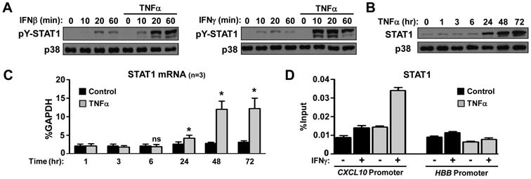
RA FLS were cultured for 3 days in the presence or absence of TNFα (10 ng/ml), which was added on the first day of culture and was not replenished (A, D). (A), On day 3 the cells were stimulated with IFNβ (1,000 U/ml) or IFNγ (100 U/ml) for 10-60 minutes and STAT1 tyrosine phosphorylation (pY) was measured by immunoblotting. The expression of STAT1 protein (B) and mRNA (C), upon TNFα time course stimulation, was measured by immunoblotting and qPCR. Values are the mean ±SEM and were normalized relative to mRNA for GAPDH. *= p<0.05 and ns=not significant, by one-tailed paired Student t test. (D), Chromatin immunoprecipitation was performed with anti-STAT1 antibody. The enrichment of immunoprecipitated STAT1 at the CXCL10 and hemoglobin B (HBB; negative control) promoters is shown as the percentage of input.