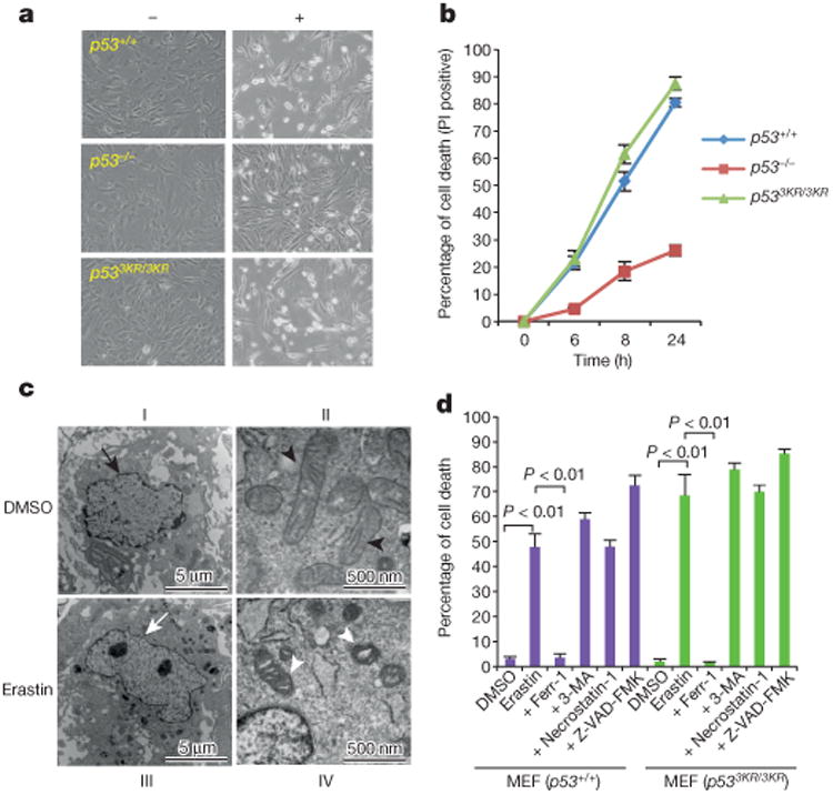
Figure 3. Roles of p53 in ferroptosis.

a, Representative phase-contrast images of MEFs treated with 4 μM erastin for 8h (magnification, ×10). b, Kinetics of cell death induced by 4 μM erastin over a 24-h period. Mean ± s.d. from two replicate experiments are shown. PI, propidium iodide. c, Wild-type MEFs were treated with dimethyl sulfoxide (DMSO) or erastin and subjected to transmission electron microscopy. Arrows, nuclei; arrow heads, mitochondria. d, MEFs were treated with erastin and specific cell death inhibitors for 8 h and the percentage of cell death was determined (error bars, s.d. from two technical replicates). 3-MA, 3-methylademine. All data are representative of three independent experiments.
