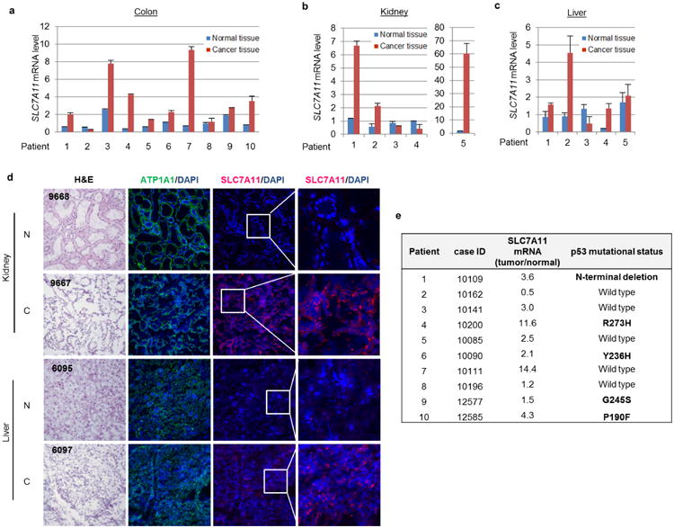
Extended Data Figure 4. SLC7A11 is overexpressed in tumours of human cancer patients.

a–c, Quantitative RT–PCR was used to determine the expression levels of SLC7A11 in paired normal and cancer tissues from colon (a), kidney (b) and liver (c); average expression levels from normal tissues were normalized to 1 in each type of cancer. Mean ± s.d. from two technical replicates are shown. d, Representative heamotoxylin and eosin (H&E) and immunofluorescence staining of SLC7A11 on frozen sections of paired patient cancer and adjacent normal tissues. Magnifcation, ×20. N, normal tissue; C, cancer tissue. Blue, DAPI; green, anti-ATP1A1; red, anti-SLC7A11. e, DNA sequencing was performed on colon cancer samples and specific mutations were identified. Independent experiments were repeated three times and representative data are shown.
