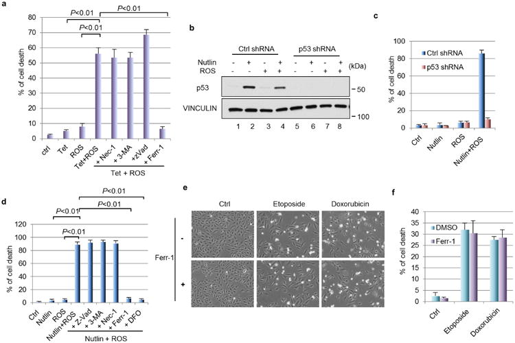
Extended Data Figure 8. Synergized ferroptosis by nutlin/ROS.

a, Percentage of cell death has shown in Fig. 6a was quantified. Mean ± s.d. from two technical replicates are shown. b, U2OS cells with stable knockdown of p53 were treated by nultin (10 μM) for 24 h followed by addition of ROS (tert-butyl hydroperoxide, 350 μM) for 4 h. Western blots were performed. c, Quantification of cell death as shown in Fig. 6c. Mean ± s.d. from two technical replicates are shown. d, U2OS cells were treated with nutlin (10 μM) for 24 h first, followed by ROS (tert-butyl hydroperoxide, 350 μM) along with indicated cell death inhibitors; cell death were quantified 24 h later. Error bars, s.d. from two technical replicates. e, U2OS cells were treated with DNA-damaging agents (etoposide, 20 μM; doxorubicin, 0.2 μg ml−1) for 48 h with or without the presence of ferr-1 (2 μM) (magnification, ×10); cell death was quantified in f with mean ± s.d. shown (n = 2 technical replicates). All data were repeated three times independently.
