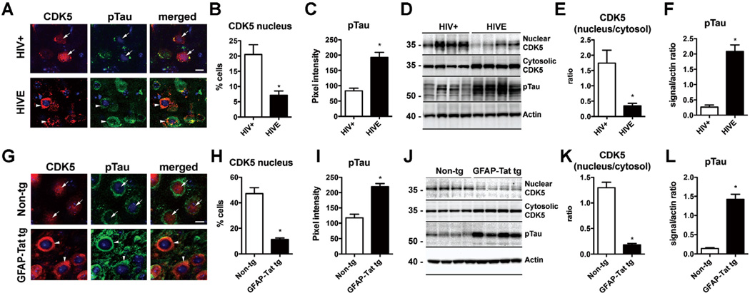
Fig. (3). Nucleo-cytoplasmic translocation of CDK5 and Tau hyperphosphorylation in HIVE and GFAP-Tat tg mice.
(A–C) Double immunolabeling and confocal microscopy for CDK5 [red] and pTau [green] in the frontal cortex of HIV+ control and HIVE patients showing increased localization of CDK5 immunostaining to the cytoplasm of HIVE cases as well as increased pTau. n=6 HIV+ and n=6 HIVE; B: p=0.0033, df=10, F=5.111; C: p=0.0002, df=10, F=3.709 by two-tailed, unpaired T-test. (D–F) Immunoblot analysis with nuclear and cytosolic fractions showing decreased CDK5 in the nuclear fraction and increased in the cytosolic fraction of HIVE cases. Levels of pTau were also increased in HIVE cases by immunoblot. n=6 HIV+ and n=6 HIVE. E: p=0.0085, df=10, F=26.69; F: p<0.0001, df=10, F=9.501 by two-tailed, unpaired T-test. Bar= 10 um. (G–I) Double immunolabeling and confocal microscopy for CDK5 [red] and pTau [green] in the cortex of non-tg and GFAP-Tat tg mice showing increased localization of CDK5 immunoreactivity to the cytoplasm of HIVE cases as well as increased pTau. n=6 non-tg and n=6 tg mice; H: p<0.0001, df=10, F=14.02; I: p<0.0001, df=10, F=1.298 (J–L) Immunoblot analysis with nuclear and cytosolic fractions showing decreased CDK5 in the nuclear fraction and increased in the cytosolic fraction of GFAP-Tat tg mice compared to non-tg mice. Levels of pTau were also increased in GFAP-Tat tg mice by immunoblot. n=6 non-tg and n=6 tg mice; K: p<0.0001, df=10, F=19.06; L: p<0.0001, df=10, F=30.13 by two-tailed, unpaired T-test. Bar= 10 um.

