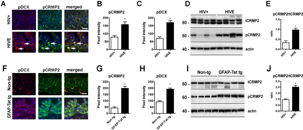
Fig. (5).
CRMP2 and DCX hyperphosphorylation in HIVE and GFAP-Tat tg mice
(A–C) Double immunolabeling and confocal microscopy for pDCX [red] and pCRMP2 [green] in the hippocampus of HIV+ control and HIVE patients showing increased pCRMP2 and pDCX in HIVE cases. n=6 HIV+ and n=6 HIVE; B: p=0.0022, df=10, F=3.19; C: p=0.0001, df=10, F=1.277 by two-tailed, unpaired T-test. (D-E) Levels of pCRMP2 were also increased in HIVE cases by immunoblot. n=6 HIV+ and n=6 HIVE; E: p=0.0006, df=10, F=1.295; by two-tailed, unpaired T-test. (F–H) Double immunolabeling and confocal microscopy for pDCX [red] and pCRMP2 [green] in the hippocampus of non-tg and tg mice showing increased pCRMP2 and pDCX in GFAP-Tat tg mice. n=6 non-tg and n=6 tg mice. G: p<0.0001, df=10, F=3.179; H: p<0.0001, df=10, F=1.133 by two-tailed, unpaired T-test. (I–J) Levels of pCRMP2 were also increased in GFAP-Tat tg mice by immunoblot. n=6 non-tg and n=6 tg mice. J: p<0.0001, df=10, F=6.875. Bar=10 um.

