
Fig. 5.
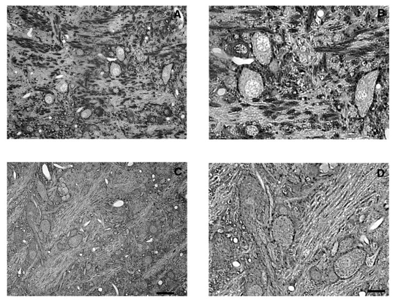
Representative photomicrographs of Gly-IR staining in the LNTB from normal hearing (A,B) and 14-day deaf (C,D) rats. B and D are enlargements of regions from A and C. In the LNTB from normal animals there is a mixture of Gly-IR and Gly-immunonegative neurons with several Gly-IR puncta surrounding each neuron. Following 14 days of deafness the number of glycine immunoreactive axosomatic puncta significantly decreases. Scale bars = 30 μm in C (applies to A,C); 15 μm in D (applies to B,D).