Fig. 9.

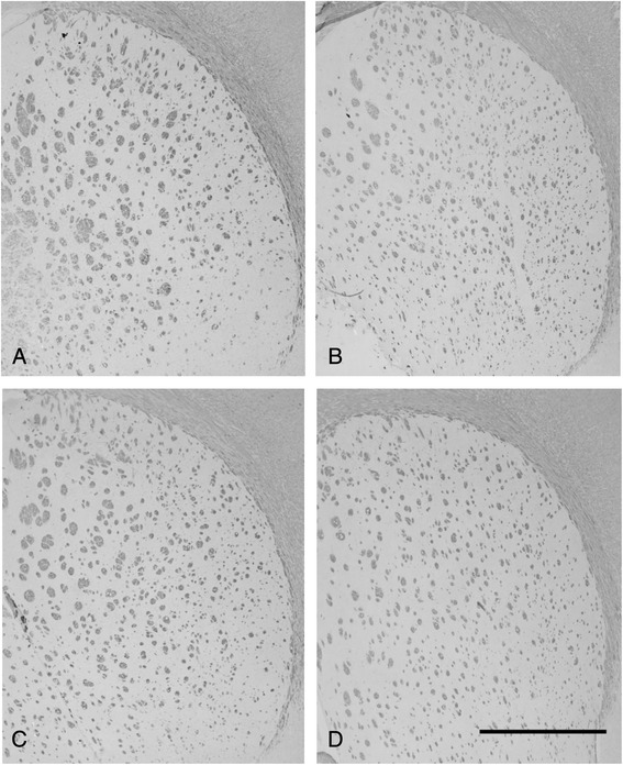
Myelin basic protein (MBP) staining at d60 of (a) N-10, (b) R-10, (c) R-6, and (d) R-16 groups. No significant differences in MBP-positive fibers could be detected. Scale bar = 1000 μm

Myelin basic protein (MBP) staining at d60 of (a) N-10, (b) R-10, (c) R-6, and (d) R-16 groups. No significant differences in MBP-positive fibers could be detected. Scale bar = 1000 μm