
Fig 2. ANKRD49 is detected primarily in spermatogonia, spermatocytes and round spermatids and localizes in the nucleus.
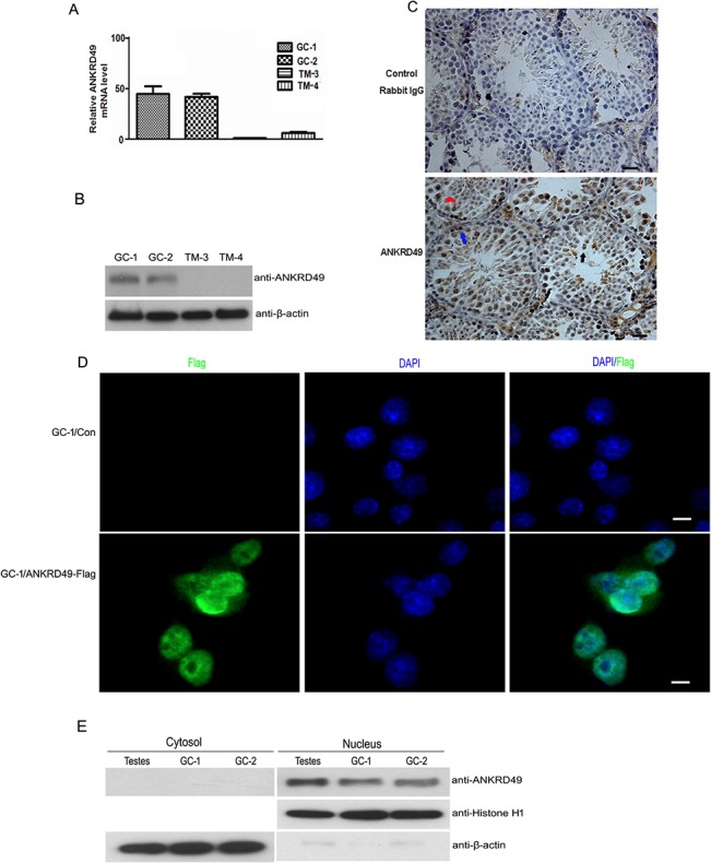
(A and B) The cell distribution of ANKRD49 mRNA and protein in four mouse testes-related cell lines were analysed by quantitative RT-PCR (A) and western blot (B). β-actin served as a loading control. (C) IHC analysis was performed with an anti-ANKRD49 polyclonal antibody (bottom panel) to detect the localization of ANKRD49 in adult mouse testes. A negative control was performed using rabbit IgG (upper panel). Blue arrow: spermatogonia; red arrow: spermatocytes; black arrow: round spermatids. Scale bars represent 50 μm. (D) ANKRD49 localizes to the nucleus. GC-1 cells which stably express ANKRD49 were subjected to IF analysis with an anti-Flag antibody followed by incubation with Alexa Fluor 488 goat anti-mouse antibody. Fluorescence signals were analysed using Confocal Laser Scanning Microscopy. Nuclei were stained with DAPI. Scale bars represent 10 μm. (E) The cytoplasm and nuclear proteins were extracted and Western blot assay was performed to detect the distribution of ANKRD49 in adult mouse testes, GC-1 and GC-2 cells. β-actin and histone H1 served as loading controls of cytoplasm and nuclear proteins, respectively. Each assay was repeated three times with similar results.