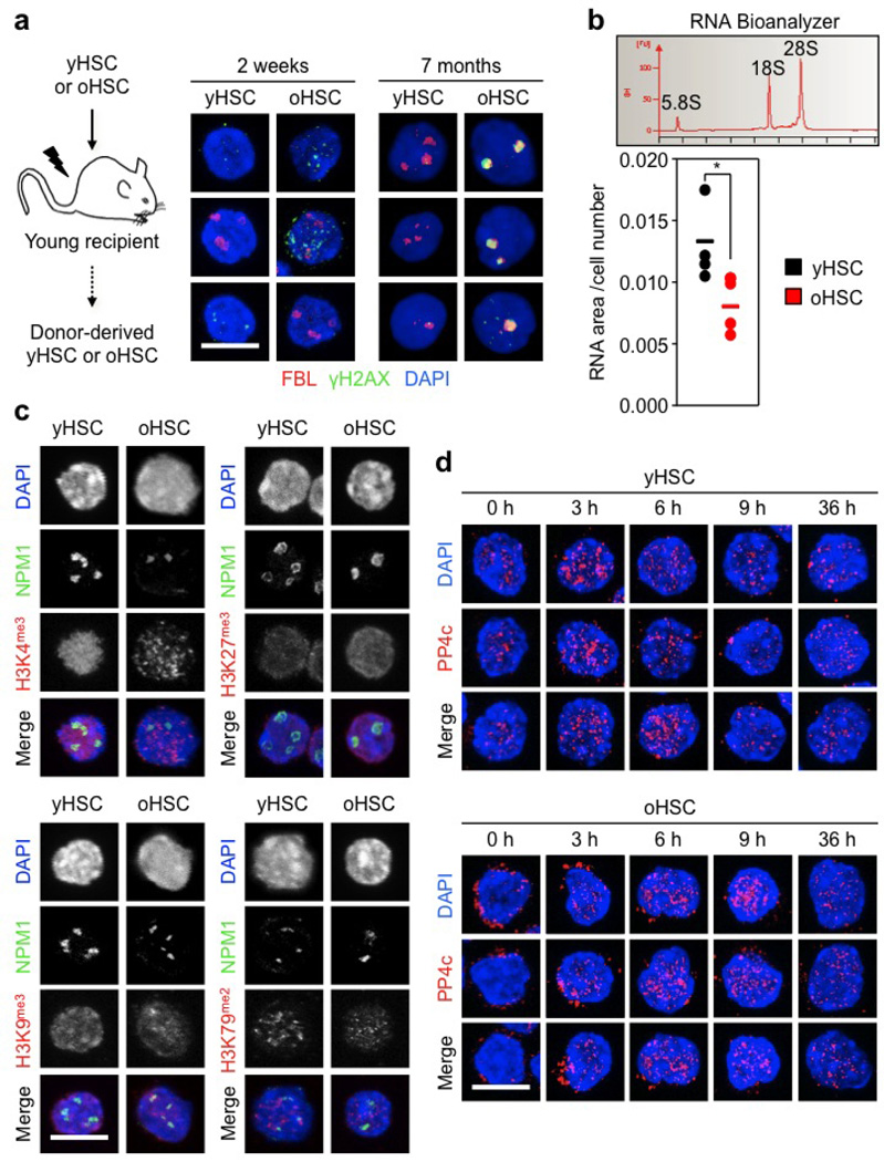
Extended Data Figure 9. Decreased ribosome biogenesis and mis-localization of the PP4c phosphatase in quiescent old HSCs.
a, Schematic and representative images of γH2AX/FBL staining in cycling and quiescent donor-derived young and old HSCs re-isolated at the indicated times after transplantation. b, RNA Bioanalyzer track showing the predominant 5.8S, 18S and 28S rRNA peaks, and quantification of RNA content in quiescent young and old HSCs (n = 4). Results are expressed as area under the curve divided by the total number of cells in each sample. Data are means ± s.d. *P ≤ 0.05. c, Representative images of NPM1 (nucleolar marker) and the indicated histone methylation mark in young and old HSCs. d, Additional images of PP4c staining in cycling young and old HSCs. Scale bars, 10 µm.

