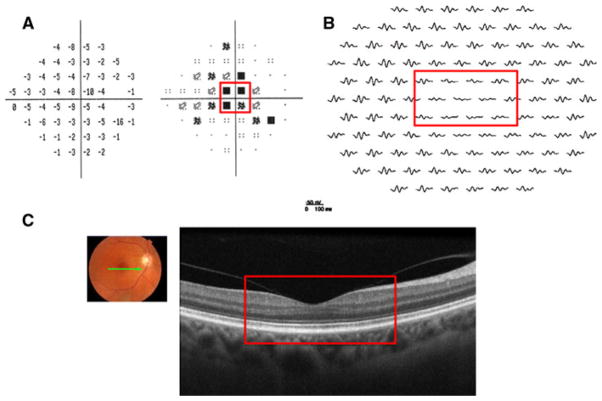
Fig. 4.
Patient 2. a 24-2 SAP demonstrates a central scotoma in the right eye (red box). b mfERG with reduced amplitude in the right eye in the retinal region corresponding to the SAP defect (red box). c fdOCT demonstrates a normal-appearing retina in the region corresponding to the VF and mfERG abnormalities (red box)

