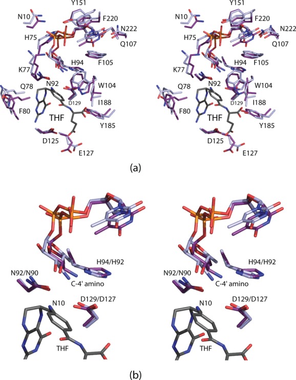
Figure 5.

Comparison of the active sites for VioF and WbtJ. The THF cofactor, as observed binding in the VioF active site, is colored in gray (a). Those residues belonging to VioF are presented in violet whereas those belonging to WbtJ are displayed in light blue. A close-up view of the region surrounding the conserved catalytic triad is presented in (b) with the same coloring scheme as described in (a). The first and second numbers in the amino acid labels correspond to VioF and WbtJ, respectively.
