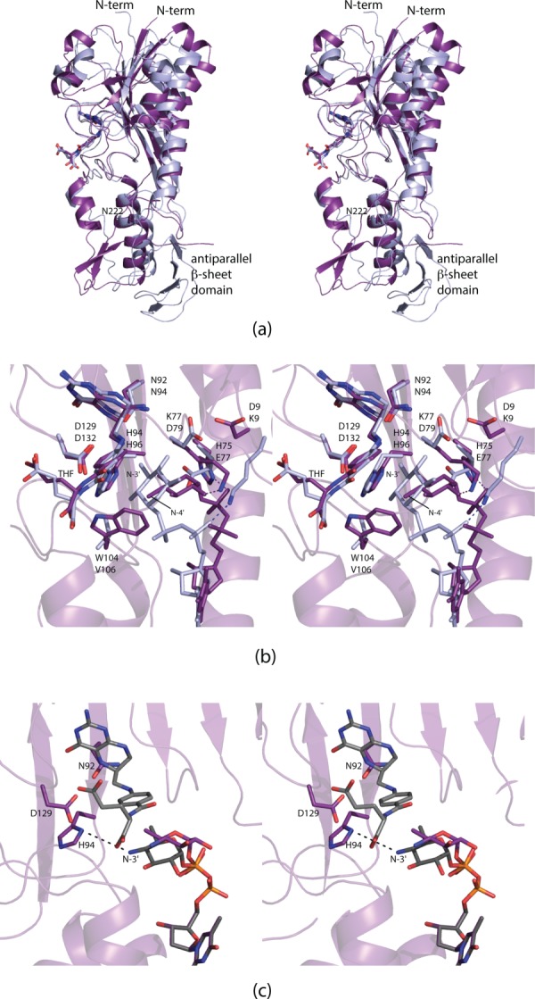
Figure 6.

Comparison of VioF with WlaRD. A superposition of the ribbon representations for the VioF (violet) and the WlaRD (light blue) subunits is presented in (a). A close-up view of their active sites is shown in (b). Note the close correspondence in the positions of the THF cofactors and the catalytic triads. It is the pyranosyl moieties of the dTDP-sugar substrates that assume markedly different locations within the active site regions. Simple rotations about the β-phosphoryl group of the dTDP-Qui4N ligand can position the C-4' amino group of the sugar to within 4.5 Å of His 94, the presumed catalytic base. This is shown in (c) where the observed conformation of the substrate pyranosyl group is highlighted in purple bonds and the “model” is displayed in gray bonds.
