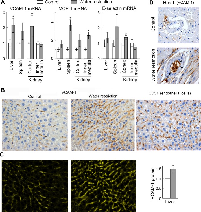
Fig 3. Water restriction activates pro-inflammatory signaling in mouse tissues.
To elevate NaCl in vivo, mice were subjected to water restriction for 9 days. To limit the amount of water, mice were fed with gel food containing 30% water and were not given any additional water. Control group mice were fed the same gel food, but had free access to water. (A) Water restriction increases VCAM-1, MCP-1 and E-selectin mRNA in several tissues. Levels of the mRNA were measured by real-time PCR. Results are presented as mean ±SEM, *P<0.05, N = 5. (B, C) Water restriction increases VCAM-1 protein in endothelial cells in the liver. (B) Representative images from immunohistochemical staining for VCAM-1 protein and for endothelial cells marker CD31 (brown) in the liver tissue sections. Similar patterns of VCAM-1 and CD31 staining are consistent with endothelial expression of VCAM-1. (C) Quantification of VCAM-1 staining shown on B. See methods section for more information about the quantification. Left panels. Images of extracted CMYK yellow channel from images shown on B. The pattern and intensity of the signal in the CMYK yellow channel corresponds to pattern and intensity to the brown diaminobenzidine (DAB) staining of VCAM-1. Right panel. Result of the quantification (mean ±SEM, *P<0.05, N = 4). (D) Representative images from immunohistochemical staining for VCAM-1 protein of heart sections. Positive staining of inner surface of coronary arteries of water restricted mice (shown by arrowheads) is consistent with endothelial expression of VCAM1. See S1 Fig for more images.

