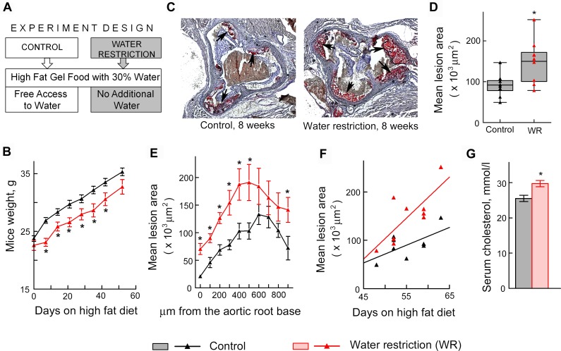
Fig 4. Water restriction accelerates atherosclerosis in ApoE-/- mice.
Mice were water restricted starting at 6 weeks of age for 7–9 weeks and atherosclerosis was analyzed in aortic root as described in methods. Data are presented as mean ±SEM, *P<0.05, N = 9. (A) Experiment design. Water restricted mice were fed with gel food made from “western” diet containing 40% of calories from fat and 30% water and were not given any additional water. Control group were fed the same gel food, but had free access to water. (B) Water restricted mice grow at the same rate as controls after transient growth retardation. (C-F) Atherosclerotic lesions are larger in water restricted mice. (C) Representative images of frozen sections through aortic root in which lipids in the atherosclerotic lesions are stained in red with Oil Red O dye (shown by arrows). (D) The graph pots mean areas of the lesions on serial sections spanning approx. 900 μm of the aortic root. The data are presented as a box-and-whisker plot (n = 9, t test, P = 0.01). (E) The graph plots mean areas of the lesions at different distances from the base of the aortic root. (F) Atherosclerotic lesions grow faster in water restricted mice as shown by steeper regression line on the scatterplot of mean lesion area vs time (see methods for more details). (G) Serum cholesterol was measured after 6 weeks on high fat diet and water restriction.

