Figure 3.
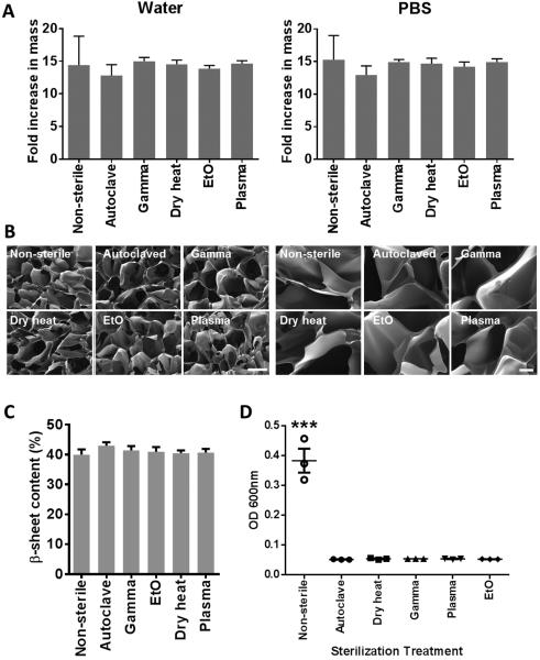
Effects of silk fibroin scaffold sterilization upon the physical characteristics of the scaffold. (A) Silk fibroin scaffold swelling in water or PBS following sterilization expressed as fold increase in mass compared to dry scaffolds, n=5. (B) Representative scanning electron microscopy images of the silk fibroin scaffolds following sterilization. The scale bar for the images on the left = 200 µm and on the right = 40 µm. (C) FTIR spectra of sterilized silk fibroin scaffolds (D) β-sheet content of sterilized silk fibroin scaffolds determined from FTIR spectra, n=4. (E) Sterility of sterilized silk fibroin scaffolds tested as bacterial growth (OD 600 nm) in Luria broth over 48h. n=3, *** p<0.001, compared to all sterilized samples.

