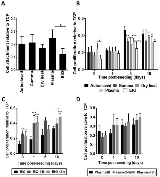
Figure 5.
Effects of silk fibroin scaffold sterilization upon cell attachment and proliferation. (A) Cell attachment after 3 h, n=5, * p<0.05. (B) Cell proliferation at 1, 5, and 10 days post seeding, n=8. p-values are calculated relative to the autoclaved samples (unless shown otherwise in the graph), * p<0.05, ** p<0.01, *** p<0.001, **** p<0.0001. (C) Cell proliferation on silk fibroin scaffolds sterilized with ethylene oxide following a 24 or 48 h incubation in PBS to leach out ethylene oxide prior to cell seeding, n=3, * p<0.05, ** p<0.01, *** p<0.001, compared to non-leached samples or as indicated in the figure. (D) Cell proliferation on silk fibroin scaffolds sterilized with H2O2 gas plasma following a 24 or 48 h incubation in PBS to leach out H2O2 prior to cell seeding, n=3 All attachment and proliferation measurements were obtained using the Alamar Blue assay.

