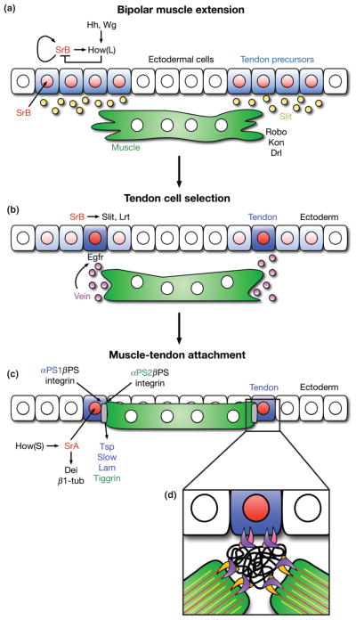
FIGURE 5.
Overview of muscle attachment to tendon cells. (a) Fields of tendon precursor cells are specified by Wingless (Wg) and Hedgehog (Hh) signaling gradients in the overlying ectoderm. All equally competent cells (blue) express StripeB (SrB, red), which auto-regulates its own expression as well as the expression of How(L), a SrB inhibitor. A balance of SrB and How(L) maintains a low level of SrB expression in all tendon precursor cells, preventing further differentiation. Tendon precursor cells secret Slit (orange), a chemoattractant for extending myofibers. The transmembrane receptor, Roundabout (Robo), on the surface of the myofiber responds to Slit secretion, and the myofiber extends filopodia toward the field of tendon precursor cells. (b) Upon Robo activation, the myofiber secretes Vein (purple), an Egfr ligand that binds to DERs on one tendon precursor cell. Interactions between Robo and Leucine-rich tendon-specific protein (Lrt), a transmembrane receptor expressed by the tendon precursor cell, solidify the attachment to the extending muscle end. (c) DER-mediated Ras signaling occurs in only the selected tendon precursor cell bound to the muscle. This leads to dedifferentiation of neighboring cells (faded blue) and upregulation of StripeA (SrA) and How(S) in the selected cell. How(S) stabilizes SrA transcripts, and SrA mediates terminal differentiation into a mature tendon cell (dark blue). A preliminary attachment between the tendon cell and the myofiber mediates the secretion of extracellular matrix proteins from both cell types (Thrombospondin (Tsp), Laminin (Lam), and Tiggrin, colored respectively), which contribute to the formation of the myotendinous junction (MTJ, gray). (d) Both cells express βPS (purple) Integrin, while the tendon cell specifically expresses αPS1 (pink) and the myofiber expresses αPS2 (orange) Integrin adhesion molecules. These transmembrane proteins heterodimerize on the surfaces of their respective cell types and bind to ECM proteins (black). α-β Integrin dimers also bind to the intracellular cytoskeletons of each cell type, forming stable connections between the tendon cell, the MTJ, and the myofiber, which can withstand the contractive forces of the mature muscle. Kon, Kon-tiki; Drl, Derailed (additional transmembrane proteins for targeting muscles to tendon cells). Dei, Delilah; β1-tub, β1-tubulin (markers of terminal tendon cell differentiation). Slow, Slowdown (secreted by tendon cell to ensure proper temporal regulation of MTJ formation). (Modified with permission from Ref 145. Copyright 2010 Company of Biologists)

