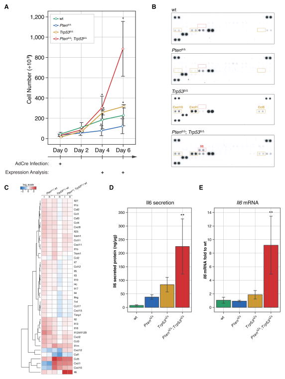
Figure 1. Il6 secretion is a hallmark of post-senescence.
(A) Combined loss of the Pten and Trp53 genes causes primary MEFs to proliferate more aggressively when compared to the single genotypes. Error bars are SD, n≥3, * p<0.05 with ANOVA, Dunnett’s post-hoc test on all genotypes vs wt at days 4 and 6.
(B) Il6 is secreted specifically after simultaneous loss of Pten and Trp53 genes. A threshold of 3-fold was used to filter up-regulated proteins specific for Trp53Δ/Δ, PtenΔ/Δ and PtenΔ/Δ; Trp53Δ/Δ cells. Ccl5 (RANTES), Cxcl1 (KC), and Cxcl10 (IP-10) proteins were up-regulated in both Trp53Δ/Δ and PtenΔ/Δ; Trp53Δ/Δ cells.
(C) Protein secretion represented as log2-transformed fold expression relative to wt and normalized to total cellular protein content. Note that there are no proteins specifically secreted in PtenΔ/Δ MEFs that exceed the 3-fold cut-off. n=3. Note that we cannot exclude stimulation of protein secretion in senescence (i.e. in the Pten-null cells) below the threshold criteria.
(D) ELISA results confirm increased expression of Il6 in conditioned medium from PtenΔ/Δ; Trp53Δ/Δ MEFs. ANOVA, Dunnett’s post-hoc test, **p<0.01, vs wt, error bars are SD, n=3.
(E) Increased transcription of Il6 is observed in PtenΔ/Δ; Trp53Δ/Δ MEFs compared to wt. ANOVA, Dunnett’s post-hoc test, **p<0.01, vs wt, error bars are SD, n=4.

