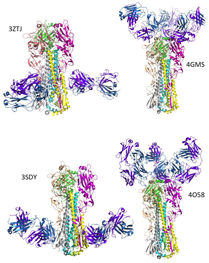
Figure 1.
Binding sites of representative broadly neutralizing antibodies on HA seen in X-ray crystal structures. The HA is shown as the trimer in approximately the same orientation in all panels. Monomer 1 colors are magenta (HA1) and yellow (HA2); monomer 2 are wheat (HA1) and gray (HA2); monomer 3 are green (HA1) and cyan (HA2). In all cases there are three antibody Fabs bound per HA trimer although the third is hidden from view in some cases. The antibody Heavy chain is colored purple and the Light chain is sky blue. The two stem-binding antibodies (F16 and CR8020, see Table 1) bind at different sites on the stem (PDB IDs 3ZTJ and 3SDY respectively). The antibodies that bind to the receptor binding region (S139/1 and F045-092) bind at different angles (PDB IDs 4GMS and 4O58 respectively). The figures were made using PyMOL Molecular Graphics System, Schrödinger, LLC.

