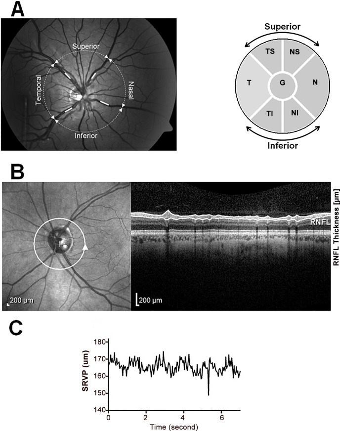
Fig 1. Sites of SRVP amplitude and RNFL thickness measurement.
A) Left- SRVP amplitude measurement at TS, NS, TI and NI sectors and Right- six sectors used for RNFL thickness measurements on OCT. B)Left-Site of RNFL measurement, Right- Cross section of retinal layers under the white circle including RNFL. C) Raw trace of SRVP.

