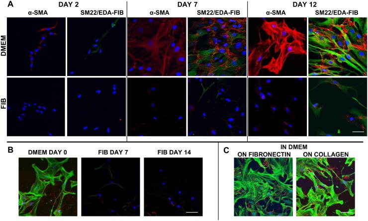
Fig 2. Confocal microscopy of cultured VICs in DMEM and FIB stained with α-SMA and doubly stained with SM22 (green) and EDA-fibronectin (red) at 2, 7 and 12 days after isolation (A).
VICs in these panels were cultured in DMEM and FIB from the point of isolation. B shows dedifferentiation of VICs from DMEM at day 0, switched to fibroblast media for 7 days (FIB day 7) and continued in fibroblast media for 14 days (FIB day 14). α-SMA is green and EDA-fibronectin is in red in panels B and C. Scale bar represents 20μm in A and 50μm in B and C.

