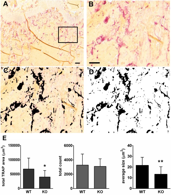
Fig 3. TRAP-positive osteoclast measurements on bone sections.
Images were obtained with a 5X objective of distal femora of WT and OCSt-KO mice at 6 weeks post-partum and TRAP-positive total area, number of TRAP-positive sites, and mean area of those sites were measured. A, A typical whole image of distal femur metaphysis, stained histochemically for TRAP (red) without counterstain. The region outlined in A is enlarged in B. C. shows the same area of the section after color matching was performed to select TRAP-positive sites in the section (now black). Finally, the images were made binary (D), leaving only TRAP-positive (black) and TRAP-negative (white) areas for analysis. Bar in A = 100 μM, bar in B = 50 μm in B, C, and D. E. The mean total TRAP-positive area per micrograph differed significantly between WT and OCSt-KO (left; *P< 0.05). The total count of TRAP-positive sites was not different between genotypes (middle); however, the mean area of each TRAP-positive sites was significantly lower in OCSt-KO (*P < 0.02). Results from 3 or 4 sections per animal, and 3 animals per genotype were pooled for analysis.

