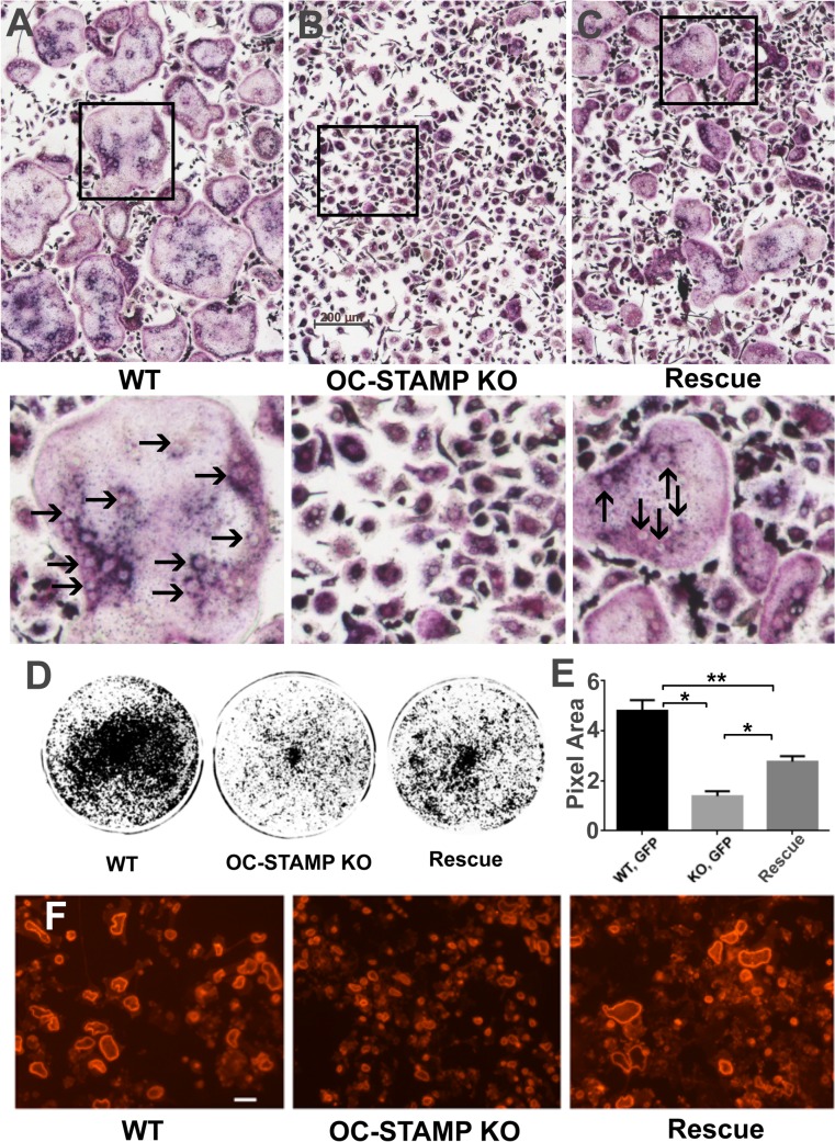
Fig 5. Rescue of OCSt-KO BMMC by lentiviral transduction.
TRAP stain shows BMMC from WT mice transduced with GFP (A: WT), from OCSt-KO mice transduced with GFP (B: OC-STAMP KO), and from OCSt-KO mice transduced with OC-STAMP:GFP (C: Rescue) following 6 days of culturing in the presence of RANKL. Many large, multinucleated osteoclasts are seen in WT cells, whereas none are present in the KO cells. Fusion is rescued by transduction with OC-STAMP. Scale bar in B = 200 μm. Boxed areas in A, B, and C upper panels are shown at higher magnification below. Some individual nuclei within multinucleated cells are indicated by arrows in the WT and Rescue panels. D. BMMC were transduced and cultured as above on HA coated plates, and the plates were scanned after 6 days of culture. Resorbed HA appears as black. E. Quantitation of resorbed area shows a roughly 3.5-fold decrease of resorption activity in KO vs. WT, whereas the rescued cells had their activity mostly restored. Mean + s.d. is shown, n = 3, *P < 0.002; **P < 0.005. F. TRITC-labeled phalloidin shows large actin rings on dentine disks in WT (left) and rescued (right) cells. KO mononuclear osteoclasts (middle) also made actin rings, but they were much smaller, consistent with small cell size. Scale bar in F = 100 μm.

