FIG 3.
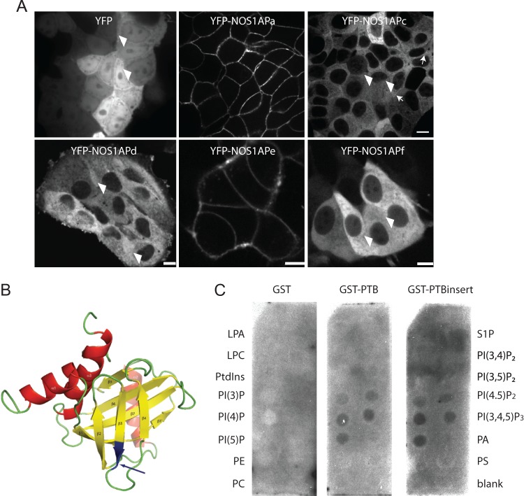
NOS1AP isoforms with different subcellular localizations. (A) Spinning-disk confocal images of MCF7 cells stably expressing YFP, YFP-NOS1APa, NOS1APc, NOS1APd, NOS1APe, and NOS1APf. Note a faint enrichment of YFP-NOS1APc at cell-cell contacts (arrows) and low levels of nuclear localization in YFP, YFP-NOS1APc, NOS1APd, and NOS1APf (arrowheads). Bar = 20 μm. (B) NMR structure model of the Drosophila NUMB PTB domain (42). The arrows point to the insert in NOS1APe positioned in the predicted surface-exposed loop in the protein. (C) A membrane covered with different phospholipids was probed with purified GST, GST-PTB, or GST-PTBinsert (the NOS1APe PTB domain containing the LLLLQ insert). LPA, lysophosphatidic acid; LPC, lysophosphatidylcholine; PE, phosphatidylethanolamine; PS, phosphatidylserine; PtdIns, phosphatidylinositol.

