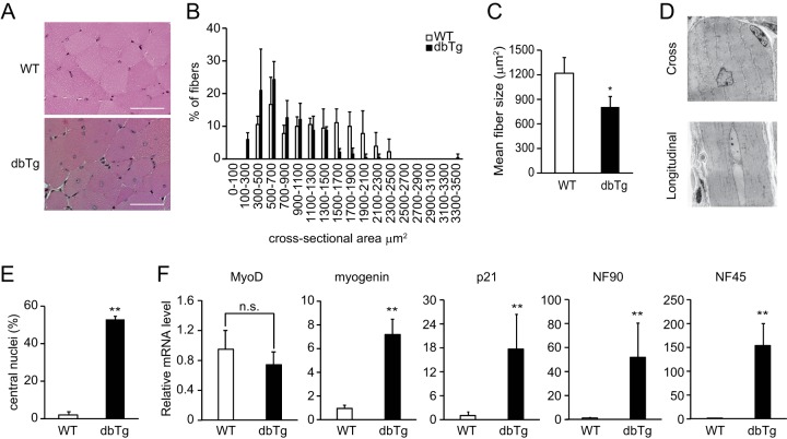
FIG 2.
Skeletal muscles of NF90-NF45 dbTg mice contain immature muscle fibers. (A) H&E staining of the cross-sectional area of quadriceps from WT and dbTg1 mice at 15 weeks of age. Bars, 50 μm. (B and C) Sizes of cross-sectional areas of quadriceps muscle fibers in WT and dbTg1 mice at 15 weeks of age were determined by using the ImageJ program. Sixty quadriceps fibers from each mouse were examined. The distribution of the cross-sectional areas (B) and the mean sizes of cross-sectional areas (C) are shown. Data shown in panels B and C are expressed as means ± standard deviations (n = 3). *, P < 0.05 compared to the WT, determined by two-tailed Student's t test. (D) Transmission electron microscopic analysis of quadriceps from dbTg1 mice at 15 weeks of age. Bars, 10 μm. (E) Number of centronuclear quadriceps fibers from WT and dbTg1 mice at 15 weeks of age. One hundred quadriceps fibers from each mouse were examined. Data are expressed as means ± standard deviations (n = 3). **, P < 0.01 compared to the WT, determined by two-tailed Student's t test. (F) Quantitative RT-PCR analysis of the expression levels of myogenic markers (MyoD, myogenin, and p21), NF90, and NF45 in quadriceps of WT and dbTg1 mice. Hypoxanthine phosphoribosyltransferase was used as an internal control. Data are expressed as means ± standard deviations (n = 4 to 5). **, P < 0.01 compared to the WT, determined by two-tailed Student's t test; n.s., not significant.

