FIG 4.
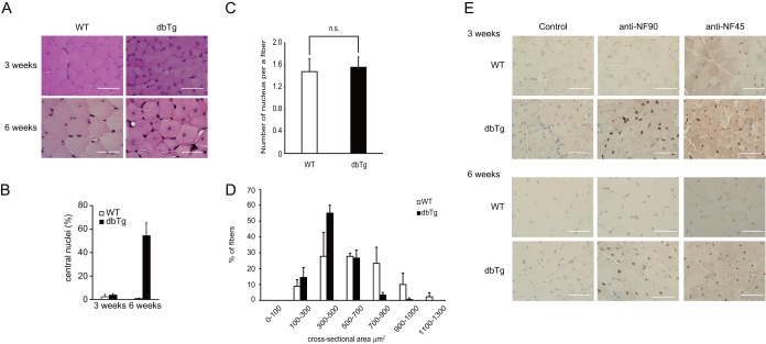
Skeletal muscles of NF90-NF45 dbTg mice exhibit centronuclear muscle fibers at 3 to 6 weeks of age. Histological analysis of the quadriceps of WT and dbTg1 mice at 3 and 6 weeks of age was performed. (A) H&E staining. Bars, 50 μm. (B) Numbers of centronuclear quadriceps fibers in WT and dbTg1 mice at 3 and 6 weeks of age. One hundred quadriceps fibers from each mouse were examined. Data are expressed as means ± standard deviations (n = 3). **, P < 0.01 compared to the WT, determined by two-tailed Student's t test. (C) Numbers of nuclei in 100 skeletal muscle fibers of WT and NF90-NF45 dbTg mice at 3 weeks of age were counted. Data are expressed as means ± standard deviations (n = 3). (D) Sizes of cross-sectional areas of quadriceps muscle fibers in WT and dbTg1 mice at 3 weeks of age were determined by using the ImageJ program. Sixty quadriceps fibers from each mouse were examined. The distribution of the cross-sectional area is shown in panel A. Data are expressed as means ± standard deviations (n = 3). (E) Immunohistochemical analysis of NF90 and NF45. (Top) mice at 3 weeks of age; (bottom) mice at 6 weeks of age. Bars, 50 μm.

