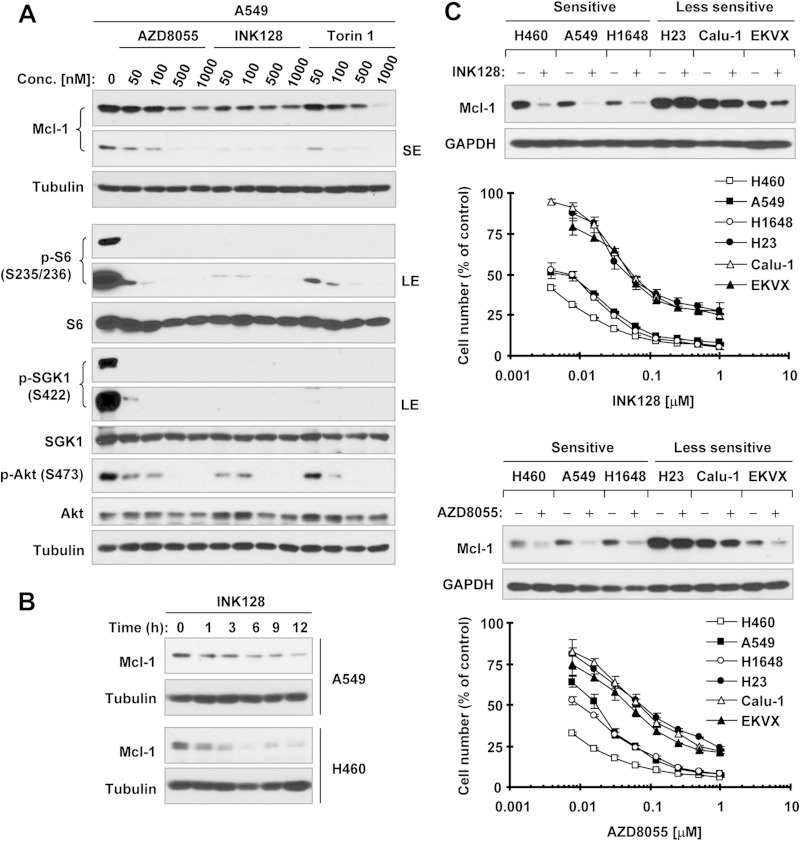
FIG 1.
TORKinibs reduce Mcl-1 levels in human NSCLC cells lines. (A) A549 cells were treated with the indicated concentrations (Conc.) of TORKinibs for 4 h. (B) The indicated cell lines were treated with 100 nM INK128 for different times as indicated. (C) The indicated NSCLC cell lines with various sensitivities to TORKinibs (lower panel) were exposed to 100 nM INK128 or AZD8055 for 4 h. After the aforementioned treatments, whole-cell protein lysates were prepared from these cell lines and subjected to Western blotting for detection of the indicated proteins. The growth curves in panel C were determined with the SRB assay after plating cells in 96-well plates and exposing them to the indicated TORKinib for 3 days. The data are means ± standard deviations of four replicate determinations. LE, long exposure; SE, short exposure; GAPDH, glyceraldehyde 3-phosphate dehydrogenase.

