FIG 3.
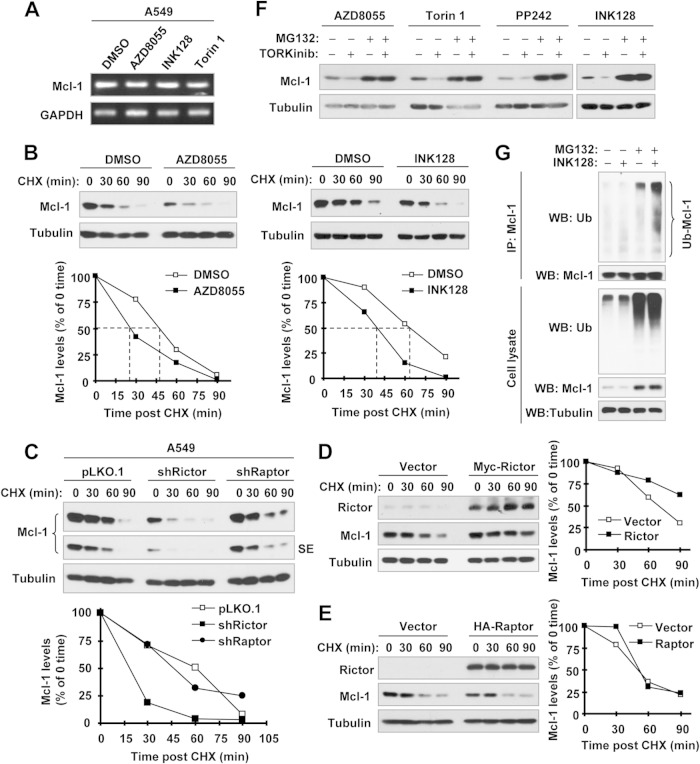
Inhibition of mTORC2 by TORKinibs (A, B, F, and G) or rictor knockdown (C) does not alter Mcl-1 mRNA levels (A) but rather promotes Mcl-1 protein degradation (B to D), whereas enforced expression of rictor but not raptor stabilizes Mcl-1 (D and E). (A) A549 cells were treated with DMSO or the indicated TORKinibs at 100 nM for 8 h. Total cellular RNA was extracted for detection of Mcl-1 mRNA levels by RT-PCR. (B) A549 cells were treated with DMSO or 100 nM INK128 or AZD8055 for 4 h. The cells were then washed with PBS three times and refed with fresh medium containing 10 μg/ml CHX. At the indicated times, the cells were harvested for preparation of whole-cell protein lysates and subsequent Western blot analysis. Protein levels were quantified with NIH ImageJ software and normalized to tubulin. (C to E) Whole-cell protein lysates were prepared from the indicated A549 transfectants exposed to 10 μg/ml CHX for different times as indicated (C) and 293T cells 48 h after transfection with myc-rictor or HA-raptor, followed by exposure to 10 μg/ml CHX for different times as indicated (D and E). The lysates were then subjected to Western blot analysis. Protein levels were quantified with NIH ImageJ software and normalized to tubulin. (F) A549 cells were pretreated with 10 μM MG132 for 1 h and then cotreated with the indicated TORKinibs (100 nM for INK128, AZD8055, and Torin 1 and 1 μM for PP242) for 4 h. The cells were harvested for preparation of whole-cell protein lysates and subsequent Western blot analysis. (G) H1299/Mcl-1 cells were transfected with HA-ubiquitin (Ub). After 48 h, the cells were pretreated with 10 μM MG132 for 40 min and then cotreated with 100 nM INK128 for another 4 h. Whole-cell protein lysates were then prepared for IP with anti-Mcl-1 antibody, followed by Western blotting (WB) with the indicated antibodies.

