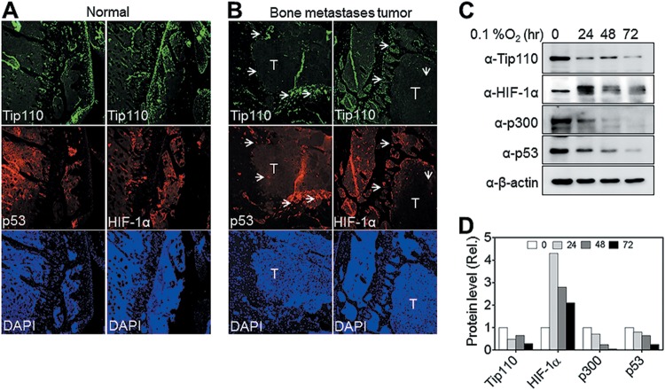
FIG 11.
Tip110, p53, and HIF-α expression in mouse bone metastases. (A and B) Mouse osteolytic bone metastasis tissues were obtained by inoculating cells of the human bone metastatic cancer melanoma cell line, 1205Lu (which contains wild-type p53), into the bones of athymic nude mice. Longitudinal sections of control (A) or metastasis (B) bone were prepared and immunostained for Tip110, p53, and HIF-1α expression. DAPI was used to visualize the nuclear DNA. T, tumor core region; arrowheads, peripheral tissues. (C and D) 1205Lu cells were cultured under severe hypoxia (0.1% O2) for the indicated lengths of time and then harvested for expression of the Tip110, p53, HIF-1α, and p300 proteins by Western blotting (C). Levels of protein expression were quantitated by using β-actin as a reference (D).

