
Figure 1.
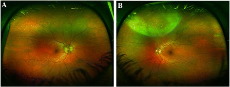
Fundus photograph of the right and left eyes. (A) Wide field color fundus photograph of the right eye: blurred optic disc margin. (B) Wide field color fundus photograph of the left eye: well-defined area of whitening involving the peripheral superonasal quadrant with slight haziness extending from that area up to the upper margin of disc and upper temporal arcade. The disc margin is ill-defined.