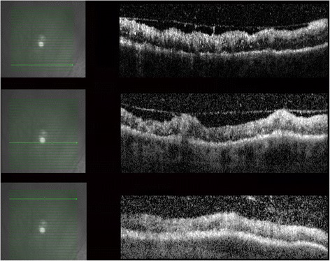
Figure 4.

OCT of the lesion: irregular retinal contour with areas of retinal elevation. The individual retinal layers could not be distinguished due to infiltration with multiple hyperreflective dots. Diffuse thickening at the retinal nerve fiber layer. Irregular vitreo-retinal interface with traction by partial PVD together with moderate hyperreflective dots in the vitreous.
