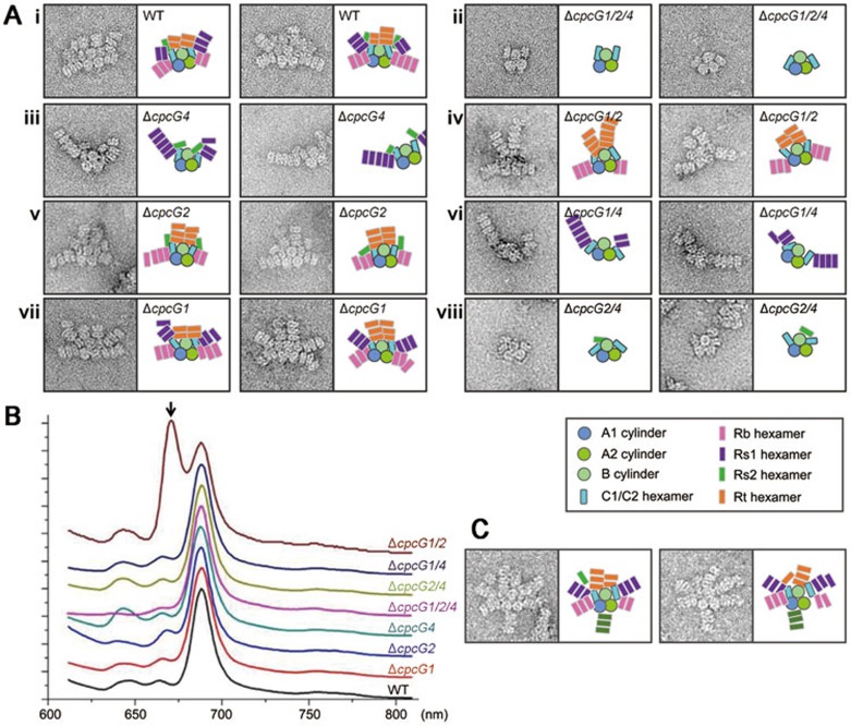
Figure 4.
Interaction of rods with the core. (A) Typical negative-stain EM images of a wild-type and mutant PBSs. The schematic models for each sample are shown to the right of the images. (B) 77K fluorescence spectra of wild-type and mutant PBSs under 590 nm excitation. The unique peak at approximately 670 nm in the ΔcpcG1/2 mutant (indicated with an arrow) may be caused by a leak of energy from the half core cylinders that cannot efficiently transfer the energy within the core. (C) Negative-stain EM images of PBSs from the ΔapcF mutant. Note that a rod is attached at the bottom of the PBS.

