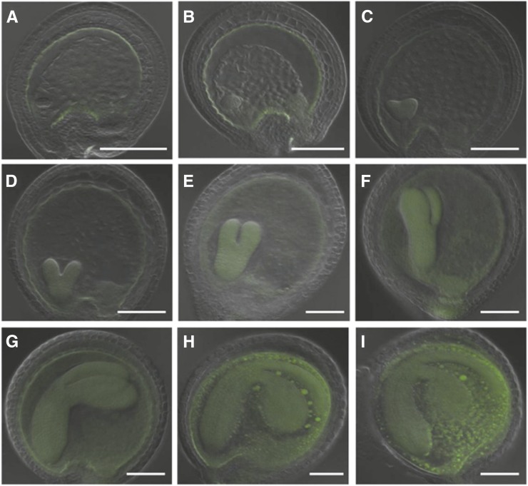
Figure 8.
Sub-Embryo Localization of CEK4-Ven in ProCEK4:CEK4-Ven cek4-1/- Using Confocal Laser Scanning Microscopy.
Embryos at the globular stage ([A] and [B]), heart stage ([C] and [D]), torpedo stage ([E] and [F]), bent cotyledon stage ([G] and [H]), and mature stage (I). Green, Ven fluorescence. Bars = 100 μm.

