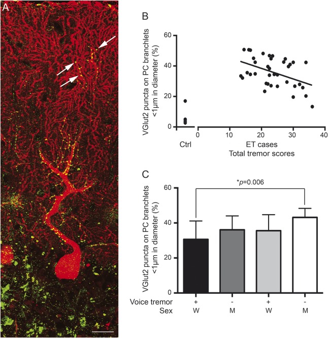
Figure. Clinicopathologic correlations of climbing fiber–Purkinje cell synaptic pathology and tremor.
(A) Dual immunofluorescence with anti-VGlut2 (Alexa 488, green) and anti-calbindin D28k antibody (Alexa 594, red) of a cerebellar section in an essential tremor (ET) case. Each Purkinje cell (PC) dendritic arbor was imaged, from the PC layers to pial surface, and reconstructed in Image J. VGlut2 puncta followed the climbing fibers and were distributed over proximal, thick PC dendrites and occasionally VGlut2 puncta localized over the distal, thin PC branchlets (arrows). Scale bar: 25 μm. (B) The percentage of VGlut2 puncta on PC branchlets <1 μm inversely correlated with the total tremor scores in ET cases. We also included data on 4 controls, collected previously.2 (C) Men without voice tremor had the highest percentage of VGlut2 puncta on PC branchlets <1 μm whereas women with voice tremor had the lowest percentage of VGlut2 puncta on PC branchlets <1 μm. M = men; W = women.

