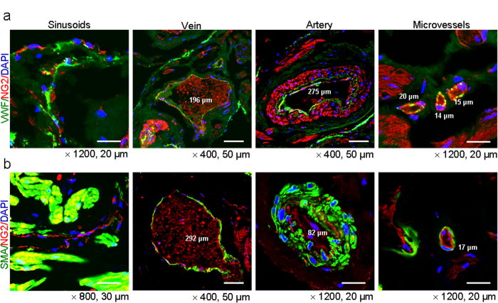
Figure 2. Localization of pericytes in the penis of congenital penile curvature patients with normal erectile function.
Merged image of transverse thin-cut (7 μm) sections. (a) Immunofluorescent double staining of penile tissue performed with antibodies against VWF (an endothelial cell marker, green) and NG2 (a pericyte marker, red). DAPI = 4,6-diamidino-2-phenylindole (a nuclei marker, blue). Scale bar = 20 μm or 50 μm. (b) Immunofluorescent souble staining of penile tissue performed with antibodies against smooth muscle α-actin (SMA, a smooth muscle cell marker, green) and NG2 (a pericyte marker, red). DAPI (a nuclei marker, blue). Scale bar = 20 μm, 30 μm, or 50 μm. Images are representative of four independent samples.

