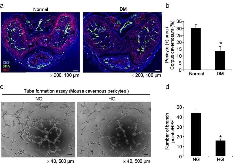
Figure 4. Decrease in pericyte content in the penis of diabetic mice.
(a) Merged images of transverse thin-cut (7 μm) sections. Immunofluorescent triple staining of penile tissue performed with antibodies against CD31 (blue), smooth muscle α-actin (SMA, green), and NG2 (red) in normal and diabetic mice. Scale bar = 100 μm and screen magnification = ×200. (b) An image analyzer was used to quantitate the NG2-immunopositive pericyte area in each group. Each bar depicts the mean ± standard deviations from n = 6 animals per group. *P < 0.01 vs. the normal group. DM = diabetes mellitus. (c) Tube formation assay in MCPs exposed to normal glucose (NG, 5 mmol) or high glucose (HG, 30 mmol) condition for 48 hours. Scale bars = 500 μm and screen magnification = ×40. (d) Number of branch points per high-power field from n = 4 wells per group. *P < 0.01 vs. the NG group.

