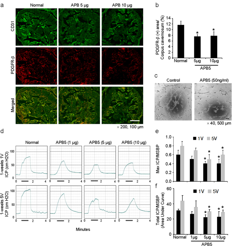
Figure 5. APB5, an anti-PGDFR-β blocking antibody, deteriorates erectile function in normal mice in vivo and pericyte function in vitro.
(a) Immunofluorescent double staining of cavernous tissue with antibodies to CD31 (green) and PDGFR-β (red) in untreated normal mice or normal mice 1 week after receiving a single intracavernous injection of PBS (20 μl) or APB5 (5 μg/20 μl or 10 μg/20 μl, respectively). Scale bar = 100 μm and screen magnification = ×200. (b) An image analyzer was used to quantitate the PDGFR-β-immunopositive area. Each bar depicts the mean ± standard deviation from n = 6 animals per group. *P < 0.01 vs. untreated normal group. (c) Tube formation assay in MCPs exposed to IgG control or APB5 (50 ng/ml) for 24 hours. Scale bars = 500 μm and screen magnification = ×40. (d) Representative intracavernous pressure (ICP) responses for untreated normal mice or normal mice stimulated at 1 week after receiving a single intracavernous injection of PBS (20 μl) or APB5 (1 μg/20 μl, 5 μg/20 μl, or 10 μg/20 μl, respectively). The cavernous nerve was stimulated at 1 and 5 V. The stimulus interval is indicated by a solid bar. (e, f) Ratios of mean maximal ICP and total ICP (area under the curve) to mean systolic blood pressure (MSBP) were calculated for each group. Each bar depicts the mean ± standard deviation from n = 6 animals per group. *P < 0.05 vs. untreated normal group. PDGFR-β, platelet-derived growth factor receptor-beta.

