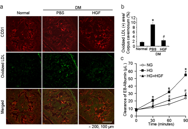
Figure 7. HGF protein transfer decreases cavernous permeability in diabetic mice in vivo and in pericytes-endothelial cell co-culture system in vitro.
(a) Immunofluorescent double staining of cavernous tissue with antibodies to CD31 (green) and oxidized LDL (red) in age-matched controls or diabetic mice 2 weeks after receiving repeated intracavernous injections of PBS (days -3 and 0; 20 μl) or HGF protein (days -3 and 0; 4.2 μg/20 μl). Scale bar = 100 μm and screen magnification = ×200. (b) An image analyzer was used to quantitate oxidized-LDL immunopositive area. Each bar depicts the mean ± standard deviation from n = 6 animals per group. *P < 0.01 vs. normal group and #P < 0.01 vs. PBS-treated diabetic group. (c) Effect of HGF on high glucose-induced pericyte-endothelial cell permeability. Pericytes and endothelial cells were cultured and treated under the following conditions: the cells exposed to normal glucose condition (NG, 5 mmol), high-glucose condition (HG, 30 mmol), and high-glucose condition (30 mmol) co-treated with rh-HGF (100 ng/ml) for 48 hours before addition of Evans blue-albumin (EBA) solution. Clearance of EBA was measured up to 90 minutes. Each bar depicts the mean ± standard deviation from six-independent experiments. *P < 0.001 vs. NG group. #P < 0.001 vs. HG group. DM = diabetes mellitus; HGF = hepatocyte growth factor; Oxidized LDL, oxidized low-density lipoprotein.

