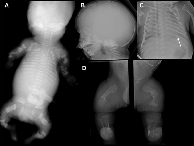
Figure 5.
Radiographs of Osteogenesis Imperfecta (OI). A. Perinatal lethal OI showing complete under mineralization of the skeleton with crumbled appendicular bone due to recurrent fractures and poor healing. B. Lateral skull of a newborn with OI. Note poor ossification of the calvarium, visualization of the anterior fontanel and wormian bones. C. A/P of the chest showing narrowness and rib fractures (arrow). D. A/P lower extremities showing poorly modeled and under-mineralized long bones, crumbled appearance due to recurrent fractures.

