
Fig. 1.
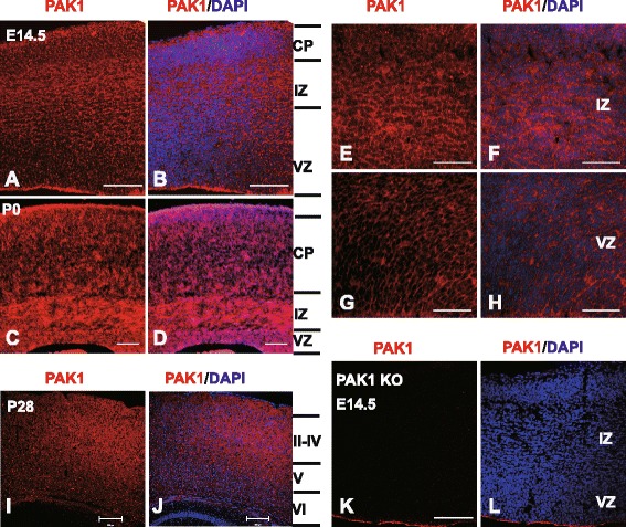
PAK1 expression in the developing mouse neocortex. Coronal brain sections of E14.5, P0 and P28 were stained with anti-PAK1 (red) and DAPI (blue). a, b PAK1/DAPI staining of WT E14.5 dorsal telencephalon. CP, cortical plate; VZ, ventricular zone; IZ, intermediate zone. c, d PAK1/DAPI staining of WT P0 neocortical sections. e-h Magnified regions of IZ and VZ areas in a and b. i, j PAK1/DAPI staining of WT P28 coronal sections. k, l PAK1/DAPI staining of PAK KO E14.5 telencephalon showing the absence of PAK1 immunostaining signals. Scale bars: 100 μm (a-d, k and l), 200 μm for (i and j), 50 μm (e-h)