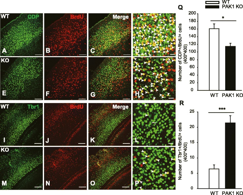
Fig. 3.

Reduced late-born pyramidal neurons in PAK1 KO P7 cortex. a–h Coronal brain sections from WT (a–d) and PAK1 KO (e–h) P7 mice were stained for the late-born neuronal marker CDP (green) and BrdU (red) injected at E14.5, showing reduced colocalization of CDP and BrdU (yellow) in PAK1 KO mice. i-p Coronal sections of the cortex of WT (i-l) and PAK1 KO (m-p) P7 mice stained for early-born neuronal markers, Tbr1 (green) and BrdU (red) injected at E14.5, showing increased colocalization of Tbr1 and BrdU. d, h and (l, p) are the magnified regions from the upper and deep layers marked by CDP and Tbr1, respectively. Arrowheads in (d, h) indicate CDP+/BrdU+ cells, in (l, p) indicate Tbr1+/BrdU+ cells. q Summary graph of CDP+/BrdU+ cells in WT and PAK1 KO mice. r Summary graph of Tbr1+/BrdU+ cells in WT and PAK1 KO mice. *p < 0.05, ***p < 0.001. Scale bars: 100 μm (a-c, e-g, i-k, m-o), 20 μm (d, h, l, p)
