Fig. 6.

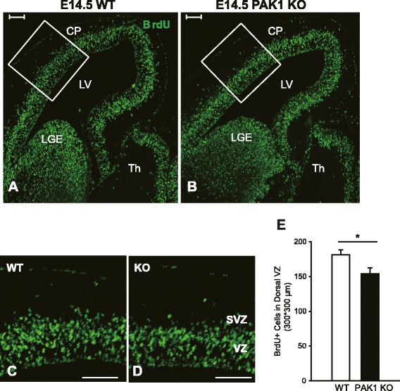
Decreased cell proliferation in the dorsal telencephalon of PAK1 KO mice. a, b Pregnant mice were labeled with BrdU at E14.5 for 2 h and the embryos were dissected and stained for BrdU. c, d Magnified views of the boxed regions in a and b. e Summary graph showing a significant reduction of BrdU positive cells in the dorsal VZ in PAK1 KO embryos compared with the WT littermates. ***p < 0.001. Scale bars: 100 μm. CP, cortical plate; LV, lateral ventricles; LGE, lateral ganglionic eminences; TH, thalamus
