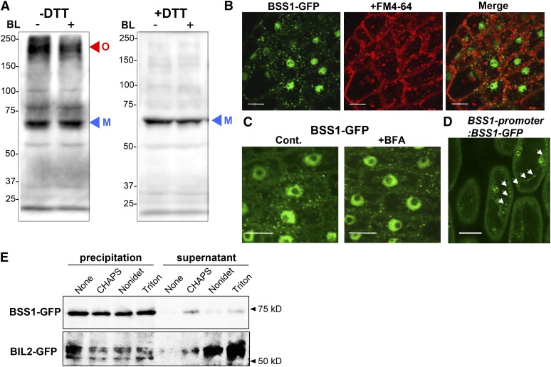
Figure 6.
Punctate Structures of BSS1/BOP1 Are a Protein Complex in the Cytoplasm.
(A) Nonreduced (−DTT) and reduced (+DTT) protein from the plant leaf was analyzed by immunoblotting using an anti-GFP antibody in transgenic plants grown on medium with Brz for 10 d. Oligomeric (O; red arrowhead) and monomeric (M; blue arrowhead) BSS1/BOP1-GFP are shown. BL induced a reduction in the amount of oligomeric and total BSS1.
(B) Root cells of BSS1/BOP1-GFP transgenic plants stained with 4 μM FM4-64 for 30 min. Bars = 10 μm.
(C) Root cells of BSS1/BOP1-GFP transgenic plants treated with 50 μM BFA for 1 h. Bars = 10 μm.
(D) Hypocotyl cells of BSS1/BOP1-promoter:BSS1/BOP1-GFP transgenic plants grown for 4 d on medium with 3 μM Brz. White arrows show BBS1/BOP1-GFP puncta. Bar = 10 μm
(E) Effects of ultracentrifugation and detergent treatment on BSS1 oligomers. Total lysates were prepared from the transgenic plants and ultracentrifuged to separate the insoluble (precipitation) and soluble (supernatant) fractions. The insoluble fractions were then treated with the indicated detergents and subjected to further ultracentrifugation. The resulting supernatants and pellets were analyzed by immunoblotting. As a control, the indicated detergent-treated BIL2-GFP (protein localized to the mitochondria) samples were centrifuged as above.

