Figure 4.
The Subcellular Localization of BES1-L in N. benthamiana and A. thaliana.
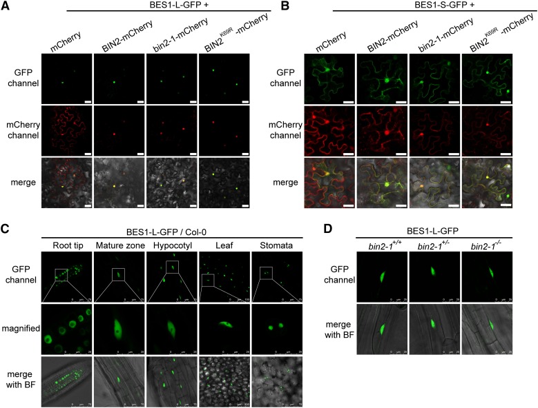
(A) and (B) The subcellular localization of BES1-L-GFP (A) or BES1-S-GFP (B) coexpressed with mCherry, BIN2-, bin2-1-, or BIN2K69R-mCherry. The indicated constructs were transformed into N. benthamiana. Bars = 40 μm.
(C) The localization of BES1-L-GFP in various tissues of A. thaliana. The 4-d-old plants were used. The middle row shows the magnified images with visible nucleolus of the corresponding regions in the upper panels. Bars are labeled in the figure.
(D) The subcellular localization of BES1-L-GFP in root mature zones of the bin2-1 gain-of-function mutant. bin2-1+/+, +/− and −/− stands for wild type, heterozygous, and homozygous mutants, respectively. Bars = 25 μm.

