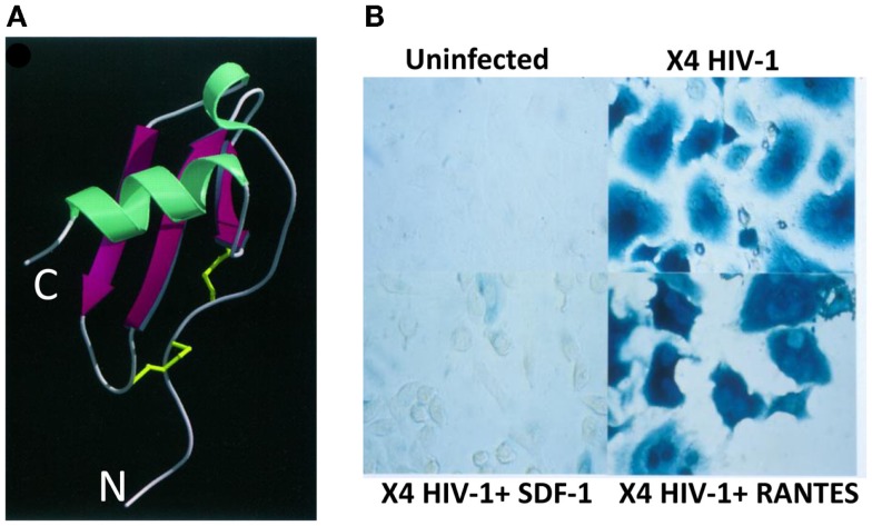
Figure 1.
Structure and mode of action of CXCL12/SDF-1 in HIV-1 infection. (A) Ribbon structure of human CXCL12/SDF-1α. The two disulfur bonds are indicated in yellow (9). N, amino-terminus; C, carboxy-terminus. (B) Human cells expressing CD4 and CXCR4 infected or not with an X4 HIV-1 isolate in the presence of SDF-1/CXCL12 or RANTES/CCL5. Infected cells are visualized by β-galactosidase staining.

