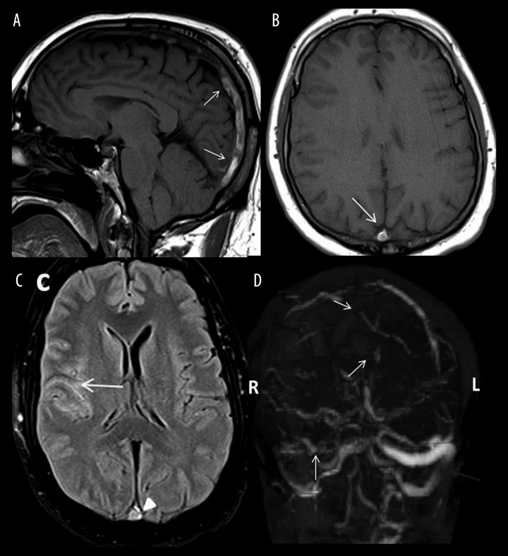
Figure 2.
Sagittal and axial T1-weighted images (A, B) showing an intrinsic bright signal within the superior sagittal sinus (arrows) with loss of a normal flow void. Axial FLAIR image (C) in the same patient showing a bright signal within the sulci (arrow) corresponding to the SAH seen on CT, along with a bright thrombus signal within the superior sagittal sinus (arrowhead). Complete non-opacification of the superior sagittal, right transverse and sigmoid sinuses (white arrows) on MR venogram (D). The left transverse sinus is patent (grey arrow).

