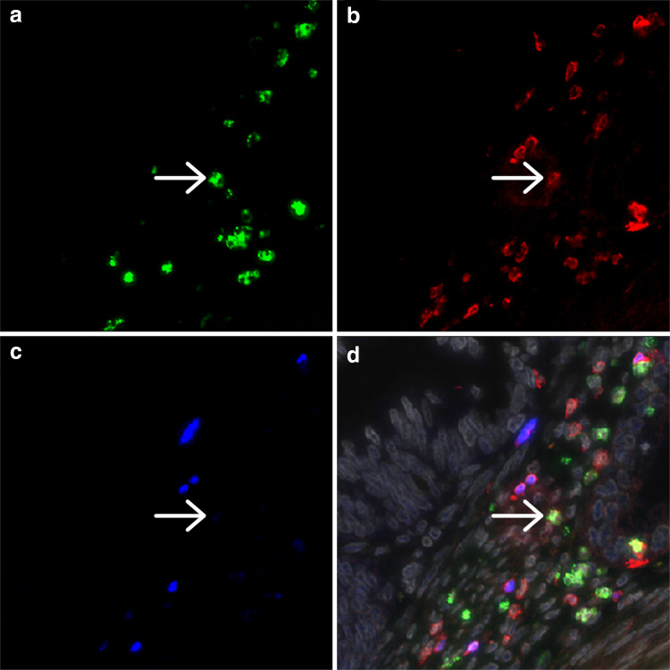
Fig. 1.
Representative image of a cervical adenocarcinoma specimen stained by triple immunofluorescence for IL-17 (a), CD3 (b) and FoxP3 (c), with the combined staining together with DAPI counterstain (gray) shown in (d). The arrow indicates a cell double positive for IL-17 and CD3. Different CD3/FoxP3 double positive cells are present

