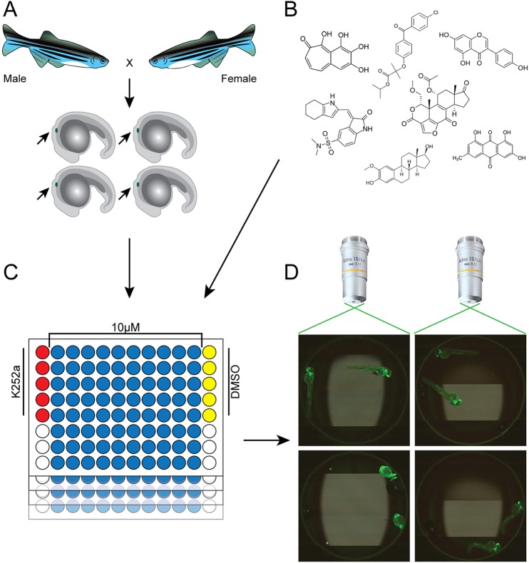
Fig. 1.
Overview of the in-vivo drug screening strategy in zebrafish. (A) The LOPAC1280, NatProd and PKIS libraries were screened for cell-migration inhibitors using 20 hpf cldnb:EGFP embryos that were GFP-positive for the migrating PLLp (marked with small arrow). (B) Compounds were transferred by ultrasonic dispenser from 384-well plates to 96-well plates so that, when 200 µl of embryo medium was added, the compounds were at a final concentration of 10 μM (NCATS, NIH). (C) Each plate contained five negative (1% DMSO) and five positive (0.01-1 μM K252a) control wells. Two embryos were placed manually into each well of the 96-well plates and PLL development was followed through 48 hpf. (D) Automated image capture of zebrafish embryos was performed using an iCys® Research Imaging Cytometer and images were scored for completion of PLLp migration.

