Fig. 7.
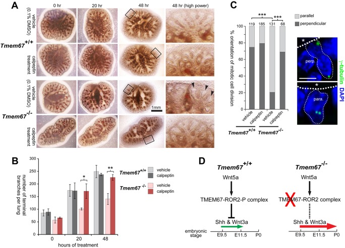
Rescue of normal embryonic lung-branching morphogenesis and polarity in mutant Tmem67−/− tissue by ex vivo treatment with the RhoA activator calpeptin. (A) Embryonic lungs (age E11.5) grown in culture for the indicated times after treatment with either vehicle control (0.1% DMSO) or calpeptin at final concentration 1 unit/ml for 3 h. Tmem67−/− lungs had abnormally dilated branches (arrowheads) surrounded by areas of condensed mesenchyme, in contrast to the fine distal branches visible in Tmem67+/+ lungs. Calpeptin treatment of mutant Tmem67−/−lungs resulted in more developed branch development and a general morphology that was similar to the wild-type lungs. Magnified insets are indicated by the black frames and shown on the right. (B) The bar graph shows the quantification of the total number of terminal branches per lung (total n=3) for each genotype and treatment condition. The statistical significance of the indicated pair-wise comparisons is *P<0.05 and **P<0.01, Student's two-tailed t-test. Error bars indicate ±s.e.m. (C) The polarity of mitotic cell division is rescued by treatment with calpeptin from predominantly parallel (para.) in mutant alveoli to predominantly perpendicular (perp.) divisions, as observed in wild-type epithelia. The statistical significance of the indicated pair-wise comparisons is ***P<0.001, chi-squared test, with the total number of cells counted in ten fields of view indicated above each bar. Representative examples of mitotic divisions, visualised by γ-tubulin (green) and indicated by the fine dotted lines, are shown on the right. Apical surfaces are highlighted by the broad dotted lines, with asterisks indicating the alveolar lumen. Scale bar: 20 μm. (D) Schematic in which signalling through the Wnt5a-TMEM67-ROR2 axis normally represses Shh and canonical Wnt (Wnt3a) signalling to moderate levels (small green arrow) between embryonic ages E9.5 and E11.5. Loss or mutation of any component in this axis (red cross) causes loss of repression (dashed line) with Shh and canonical Wnt pathway de-regulation and ectopic expression of Shh at later gestation ages (large red arrow). This contributes to pulmonary hypoplasia with condensed mesenchyme and impaired development of the alveolar system in the ciliopathy disease state.

